
_1.jpg)
|
|
Characterization and Application of Nanomaterials (CAN) is an open access peer-reviewed journal allowing maximum visibility of articles published in it as they are available to a wide, global audience. We are interested in the scientific topics from all fields of nano. CAN provides a forum to share scholarly practice to advance the use of nanomaterials in the context of scientific application. CAN publishes original research articles, review articles, editorials, case reports, brief commentaries, perspectives, etc. Examples of relevant topics include but are not limited to:
Journal Abbreviation: Charact. Appl. Nanomater. |
Online Submissions
Registration and login are required to submit items online and to check the status of current submissions.
Already have a Username/Password for Characterization and Application of Nanomaterials?
GO TO LOGIN
Need a Username/Password?
GO TO REGISTRATION
Submission Preparation Checklist
As part of the submission process, authors are required to check off their submission's compliance with all of the following items, and submissions may be returned to authors that do not adhere to these guidelines.
- The submission has not been previously published, nor is it under the consideration of another journal (or an explanation has been provided in Comments to the Editor).
- The submission file is in Microsoft Word format.
- Where available, URLs for the references have been provided.
- The text adheres to the stylistic and bibliographic requirements outlined in the Author Guidelines, which is found in About the Journal.
- If submitting to a peer-reviewed section of the journal, the instructions in Ensuring a Blind Review have been followed.
Privacy Statement
EnPress Publisher respects and strives to protect the privacy of its users and visitors. Hence, users and visitors are encouraged to read EnPress Publisher’s privacy policy regarding the usage and handling of user information.
(1) User information
Names and email addresses entered in all EnPress Publisher’s journal sites will be used exclusively for the stated purposes of the journals and will not be made available for any other purpose or to any other party. For submission and peer review, users should register an account for further procedures, including but not limited to name, email, address, interests, affiliation, and postcode, as editors need the information to complete in-house processes (e.g., processing a manuscript).
When users visit the publisher's website, information about the visit is saved in web logs (e.g., device, IP address, time of visit, etc.), which are only used to help improve the structure and content of the website.
(2) User rights
Users have the right to register or update their personal information and contact the publisher to cancel/delete their account if required.
(3) Third-party link
EnPress Publisher is not responsible for private information obtained by third-party websites when users log in via a pop-up screen from third-party software installed on their computer.
When users visit third-party platforms (e.g., LinkedIn, Twitter, COPE, etc.) through hyperlinks from EnPress Publisher’s journal websites, the privacy policy follows the policies of the third-party platforms.
(4) Queries or contact
For any queries about EnPress Publisher’s privacy policy, please contact the editorial office at editorial@enpress-publisher.com.
Article Processing Charges (APCs)
Characterization and Application of Nanomaterials is an Open Access Journal under EnPress Publisher. All articles published in Characterization and Application of Nanomaterials are accessible electronically from the journal website without commencing any kind of payment. In order to ensure contents are freely available and maintain publishing quality, Article Process Charges (APCs) are applicable to all authors who wish to submit their articles to the journal to cover the cost incurred in processing the manuscripts. Such cost will cover the peer-review, copyediting, typesetting, publishing, content depositing and archiving processes. Those charges are applicable only to authors who have their manuscript successfully accepted after peer-review.
| Journal Title | APCs |
|---|---|
| Characterization and Application of Nanomaterials | $1000 |
We encourage authors to publish their papers with us and don’t wish the cost of article processing fees to be a barrier especially to authors from the low and lower middle income countries/regions. A range of discounts or waivers are offered to authors who are unable to pay our publication processing fees. Authors can write in to apply for a waiver and requests will be considered on a case-by-case basis.
*Article No. is mandatory for payment and it can be found on the acceptance letter issued by the Editorial Office. Payment without indicating Article No. will result in processing problem and delay in article processing. Please note that payments will be processed in USD. You can make payment through Masters, Visa or UnionPay card.
Vol 8, No 3 (2025)
Table of Contents
 Open Access
Open Access
Open Access
Open Access
Open Access
The tunable conduction of coumarin-based composites has attracted considerable attention in a wide range of applications due to their unique chemical structures and fascinating properties. The incorporation of graphene oxide (GO) further enhances coumarin properties, including strong fluorescence, reversible photodimerization, and good thermal stability, expanding their potential use in advanced technological applications. This review describes the developmental evolution from GO, GO-polymer, and coumarin-based polymer to the coumarin-GO composite, concerning their synthesis, characterization, unique properties, and wide applications. We especially highlight the outstanding progress in the synthesis and structural characteristics along with their physical and chemical properties. Therefore, understanding their structure-property relations is very important to acquire scientific and technological information for developing the advanced materials with interesting performance in optoelectronic and energy applications as well as in the biomedical field. Given the expertise of influenced factors (e.g., dispersion quality, functionalization, and loading level) on the overall extent of enhancement, future research directions include optimizing coumarin-GO composites by varying the nanofiller types and coumarin compositions, which could significantly promote the development of next-generation polymer composites for specific applications.
Open Access
In this review are developed insights from the current research work to develop the concept of functional materials. This is understood as real modified substrates for varied applications. So, functional and modified substrates focused on nanoarchitectures, microcapsules, and devices for new nanotechnologies highlighting life sciences applications were revised. In this context, different types of concepts to proofs of concepts of new materials are shown to develop desired functions. Thus, it was shown that varied chemicals, emitters, pharmacophores, and controlled nano-chemistry were used for the design of nanoplatforms to further increase the sizes of materials. In this regard, the prototyping of materials was discussed, affording how to afford the challenge in the design and fabrication of new materials. Thus, the concept of optical active materials and the generation of a targeted signal through the substrate were developed. Moreover, advanced concepts were introduced, such as the multimodal energy approach by tuning optical coupling from molecules to the nanoscale within complex matter composites. These approaches were based on the confinement of specific optical matter, considering molecular spectroscopics and nano-optics, from where the new concept nominated as metamaterials was generated. In this manner, fundamental and applied research by the design of hierarchical bottom-up materials, controlling molecules towards nanoplatforms and modified substrates, was proposed. Therefore, varied accurate length scales and dimensions were controlled. Finally, it showed proofs of concepts and applications of implantable, portable, and wearable devices from cutting-edge knowledge to the next generation of devices and miniaturized instrumentation.
Open Access
Open Access
Announcements
Characterization and Application of Nanomaterials accepted for inclusion in Scopus |
|
We are thrilled to announce that Characterization and Application of Nanomaterials (CAN) has been officially accepted for inclusion in Scopus! This milestone reflects our commitment to advancing high-quality research and fostering global scholarly exchange. |
|
| Posted: 2025-08-13 | More... |
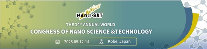
CAN supports the 14th Annual World Congress of Nano Science and Technology as the journal partner! |
|
|
|
| Posted: 2025-04-27 | More... |
CAN cooperates with 4th International Conference on Advanced Nanomaterials and Nanotechnology as a supported journal. |
|
 Scisynopsis warmly invites attendees from all over the world to attend the "4th International Conference on Advanced Nanomaterials and Nanotechnology" in H10 Roma Città, Rome, Italy, from June 12 to 13, 2025. |
|
| Posted: 2025-04-25 | More... |
| More Announcements... |

This site is licensed under a Creative Commons Attribution 4.0 International License.