


|
Journal Abbreviation: Imaging. Radiat. Res. |
Imaging and Radiation Research (IRR, ISSN: 2578-1618) is an international journal dedicated to advancing the field of medical imaging, radiation sciences, and their applications in health and disease. IRR is open to a broad range of subjects, spanning medical science, surgical practices, biomedical engineering, biology, materials science, environmental science, and related branches of physics and chemistry. It welcomes original research contributions, including laboratory-based studies, modeling, field tests, case reports, reviews, and significant applications of imaging technology and radiation-related analysis. The journal covers all aspects of imaging technology and analysis methods, Radiation Biology & Radiation Physics, including but not limited to:
|
Online Submissions
Registration and login are required to submit items online and to check the status of current submissions.
Already have a Username/Password for Imaging and Radiation Research?
GO TO LOGIN
Need a Username/Password?
GO TO REGISTRATION
Submission Preparation Checklist
As part of the submission process, authors are required to check off their submission's compliance with all of the following items, and submissions may be returned to authors that do not adhere to these guidelines.
- The submission has not been previously published, nor is it under another journal's consideration (or an explanation has been provided in Comments to the Editor).
- The submission file is in Microsoft Word format.
- Where available, URLs for the references have been provided.
- The text adheres to the stylistic and bibliographic requirements outlined in the Author Guidelines, which is found in About the Journal.
- If submitting to a peer-reviewed section of the journal, the instructions in Ensuring a Blind Review have been followed.
Privacy Statement
EnPress Publisher respects and strives to protect the privacy of its users and visitors. Hence, users and visitors are encouraged to read EnPress Publisher’s privacy policy regarding the usage and handling of user information.
(1) User information
Names and email addresses entered in all EnPress Publisher’s journal sites will be used exclusively for the stated purposes of the journals and will not be made available for any other purpose or to any other party. For submission and peer review, users should register an account for further procedures, including but not limited to name, email, address, interests, affiliation, and postcode, as editors need the information to complete in-house processes (e.g., processing a manuscript).
When users visit the publisher's website, information about the visit is saved in web logs (e.g., device, IP address, time of visit, etc.), which are only used to help improve the structure and content of the website.
(2) User rights
Users have the right to register or update their personal information and contact the publisher to cancel/delete their account if required.
(3) Third-party link
EnPress Publisher is not responsible for private information obtained by third-party websites when users log in via a pop-up screen from third-party software installed on their computer.
When users visit third-party platforms (e.g., LinkedIn, Twitter, COPE, etc.) through hyperlinks from EnPress Publisher’s journal websites, the privacy policy follows the policies of the third-party platforms.
(4) Queries or contact
For any queries about EnPress Publisher’s privacy policy, please contact the editorial office at editorial@enpress-publisher.com.
Article Processing Charges (APCs)
Imaging and Radiation Research is an Open Access Journal under EnPress Publisher. All articles published in Imaging and Radiation Research are accessible electronically from the journal website without commencing any kind of payment. In order to ensure contents are freely available and maintain publishing quality, Article Process Charges (APCs) are applicable to all authors who wish to submit their articles to the journal to cover the cost incurred in processing the manuscripts. Such cost will cover the peer-review, copyediting, typesetting, publishing, content depositing and archiving processes. Those charges are applicable only to authors who have their manuscript successfully accepted after peer-review.
| Journal Title | APCs |
|---|---|
| Imaging and Radiation Research | $300 |
We encourage authors to publish their papers with us and don’t wish the cost of article processing fees to be a barrier especially to authors from the low and lower middle income countries/regions. A range of discounts or waivers are offered to authors who are unable to pay our publication processing fees. Authors can write in to apply for a waiver and requests will be considered on a case-by-case basis.
APCs Payment
Payments for APC of this journal can be made through our online PayPal payment gateway. Enter the Article No. into the below textbox and select "Pay Now" to proceed with payment.
*Article No. is mandatory for payment and it can be found on the acceptance letter issued by the Editorial Office. Payment without indicating Article No. will result in processing problem and delay in article processing. Please note that payments will be processed in USD. You can make payment through Masters, Visa or UnionPay card.
Vol 8, No 1 (2025)
Table of Contents
 Open Access
Instant and accurate evaluation of drug resistance in tumors before and during chemotherapy is important for patients with advanced colon cancer and is beneficial for prolonging their progression-free survival time. Here, the possible biomarkers that reflect the drug resistance of colon cancer were investigated using proton magnetic resonance spectroscopy (1H-MRS) in vivo. SW480[5-fluorouracil(5-FU)-responsive] and SW480/5-FU (5-FU-resistant) xenograft models were generated and subjected to in vivo 1H-MRS examinations when the maximum tumor diameter reached 1–1.5 cm. The areas under the peaks for metabolites, including choline (Cho), lactate (Lac), glutamine/glutamate (Glx), and myo-inositol (Ins)/creatine (Cr) in the tumors, were analyzed between two groups. The resistance-related protein expression, cell morphology, necrosis, apoptosis, and cell survival of these tumor specimens were assessed. The content for tCho, Lac, Glx, and Ins/Cr in the tumors of the SW480 group was significantly lower than that of the SW480/5-FU group (P < 0.05). While there was no significant difference in the degree of necrosis and apoptosis rate of tumor cells between the two groups (P > 0.05), the tumor cells of the SW480/5-FU showed a higher cell density and larger nuclei. The expression levels of resistance-related proteins (P-gp, MPR1, PKC) in the SW480 group were lower than those in the SW480/5-FU group (P < 0.01). The survival rate of 5-FU-resistant colon cancer cells was significantly higher than that of 5-FU-responsive ones at 5-FU concentrations greater than 2.5 μg/mL (P < 0.05). These results suggest that alterations in tCho, Lac, Glx1, Glx2, and Ins/Cr detected by 1H-MRS may be used for monitoring tumor resistance to 5-FU in vivo.
Open Access
Abstract: Retinal disorders, such as diabetic retinopathy, glaucoma, macular edema, and vein occlusions, are significant contributors to global vision impairment. These conditions frequently remain symptomless until patients suffer severe vision deterioration, underscoring the critical importance of early diagnosis. Fundus images serve as a valuable resource for identifying the initial indicators of these ailments, particularly by examining various characteristics of retinal blood vessels, such as their length, width, tortuosity, and branching patterns. Traditionally, healthcare practitioners often rely on manual retinal vessel segmentation, a process that is both time-consuming and intricate, demanding specialized expertise. However, this approach poses a notable challenge since its precision and consistency heavily rely on the availability of highly skilled professionals. To surmount these challenges, there is an urgent demand for an automatic and efficient method for retinal vessel segmentation and classification employing computer vision techniques, which form the foundation of biomedical imaging. Numerous researchers have put forth techniques for blood vessel segmentation, broadly categorized into machine learning, filtering-based, and model-based methods. Machine learning methods categorize pixels as either vessels or non-vessels, employing classifiers trained on hand-annotated images. Subsequently, these techniques extract features using 7D feature vectors and apply neural network classification. Additional post-processing steps are used to bridge gaps and eliminate isolated pixels. On the other hand, filtering-based approaches employ morphological operators within morphological image processing, capitalizing on predefined shapes to filter out objects from the background. However, this technique often treats larger blood vessels as cohesive structures. Model-based methods leverage vessel models to identify retinal blood vessels, but they are sensitive to parameter selection, necessitating careful choices to simultaneously detect thin and large vessels effectively. Our proposed research endeavors to conduct a thorough and empirical evaluation of the effectiveness of automated segmentation and classification techniques for identifying eye-related diseases, particularly diabetic retinopathy and glaucoma. This evaluation will involve various retinal image datasets, including DRIVE, REVIEW, STARE, HRF, and DRION. The methodologies under consideration encompass machine learning, filtering-based, and model-based approaches, with performance assessment based on a range of metrics, including true positive rate (TPR), true negative rate (TNR), positive predictive value (PPV), negative predictive value (NPV), false discovery rate (FDR), Matthews's correlation coefficient (MCC), and accuracy (ACC). The primary objective of this research is to scrutinize, assess, and compare the design and performance of different segmentation and classification techniques, encompassing both supervised and unsupervised learning methods. To attain this objective, we will refine existing techniques and develop new ones, ensuring a more streamlined and computationally efficient approach.
Open Access
Objective: Standardizing image acquisition protocols and image quality across cameras is an important need in imaging, in particular in multi-center clinical trials and the use of image analysis and machine learning algorithms. The objective of this study was to examine the effect of ordered subset expectation maximization (OSEM) reconstruction parameters on the quantitative image quality of cardiac perfusion SPECT images in different typical SPECT cameras and therefore assess the need to change the parameter values across cameras.
Methods: The analysis was carried out by comparing the defect contrast-to-noise ratio (CNR) at 12 OSEM subset-iteration combinations. Eight frames were reconstructed using the SIMIND Monte Carlo Simulation package. An activity of 370 MBq (10mCi) and projection acquisition interval of 20 seconds per projection were used. Attenuation (AC) and scatter corrections (SC) were performed in this study for all images.
Results: The 16-2 subset-iteration combination yielded the highest CNR and defect contrast values for both cameras. The difference between CNR values for two cameras was found to be close to 5%.
Conclusions: Monte Carlo simulations can be useful to investigate how quantitative image quality behaves with respect to reconstruction parameters and correction algorithms in a controlled environment. In this study, the use of different camera brands did not seem to significantly affect the lesion detectability. Further simulations with more extended range of parameters and camera brands may be conducted in the future to quantify further the variability between different brands of cameras.
Open Access
This document outlines the advancements in AI- accelerated frame generation utilizing Neural Processing Units (NPU) in mobile devices. The integration of NPU technology enhances the processing efficiency of mobile graphics, enabling real-time frame generation that significantly improves video and image quality. By leveraging specialized hardware designed for AI computations, the system reduces latency and optimizes power consumption, making it ideal for demanding applications such as gaming and augmented reality. This paper discusses the underlying architecture of NPUs, their role in accelerating frame generation, and the potential impacts on user experience in mobile environments. The findings illustrate how NPU-driven solutions can transform mobile graphics, offering a more immersive and responsive experience while efficiently managing resources.
Announcements
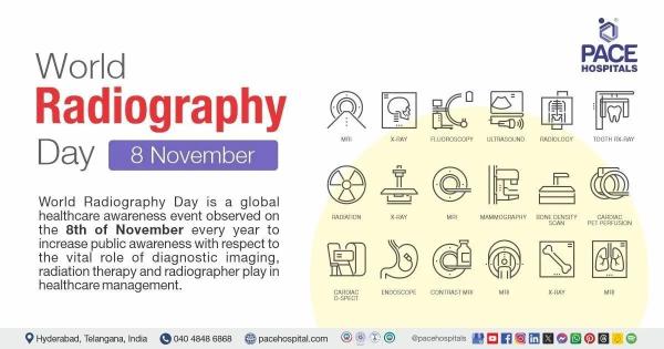
World Radiography Day 8 November 2024 - Importance, Theme & History |
|
 @ww.pacehospital.com |
|
| Posted: 2024-11-08 | More... |
Fourth Committee Considers Effects of Atomic Radiation, Reaffirms Commitment to UN Scientific Committee, Hears Reports on Information, Peacekeeping |
|
Reaffirming the important work of the United Nations Scientific Committee on the Effects of Atomic Radiation, the Fourth Committee (Special Political and Decolonization) today approved without a vote a resolution concerning that body, as speakers underscored the imperative of concerted and apolitical scientific work on this matter while also considering how to improve their working methods. |
|
| Posted: 2024-10-22 | More... |
Requirements for Documentation on Human and Animal Research |
|
| For articles submitted to the journal Imaging and Radiation Research that involve human or animal research, authors are required to provide the following documentation based on the specific circumstances: | |
| Posted: 2024-08-18 | More... |
| More Announcements... |

This site is licensed under a Creative Commons Attribution 4.0 International License.